

After researching in the anatomical, radiologicaland histological aspects of more than 30 human placentas, our research grouphas found many close resemblances between the chorionic plate vessels and thehuman brain vessels. Since the placental cells are still viable soon after delivery, it would be possible to covert such otherwise due for disposable organ into an ex-vivo vascular model for testing of newly designed neurointerventional devices. The model is easy to set up and requires no sacrifice of animal. We hope that our suggested model can be repeated in other research institute and adopting it as a useful tool for testing new design and training for young neurointerventionalist.
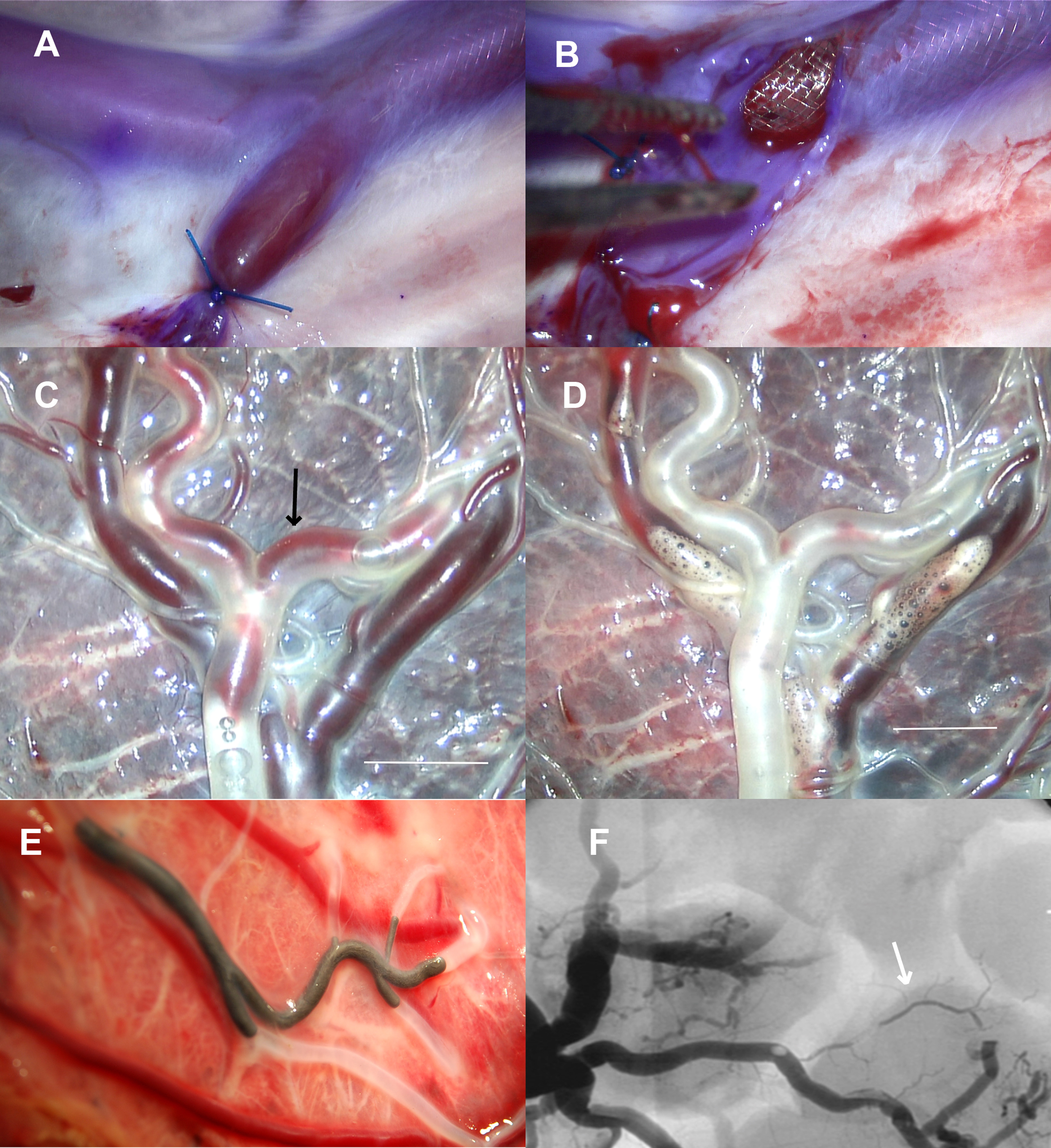
Human placentas are obtained by written consent and request for donation of organ for research from themother before delivery of the baby. 18 placentas were divided into varioussmall groups for simulation of neurointervention procedures. Pathological conditions such as acute thrombotic stroke, arterio-venous malformation and small saccular aneurysm can be mimicked by modification of the chorionic plate vessels. Thus treatment methods such as thrombolysis, pushing of glue, embolization of coils and deployment of flow diverter can be studied either under directvision with surgical microscope or digital subtraction angiography. The outcomes are found to closely resemble clinical situations and potential interventional complications explored. The vascular model we described can be further expanded in many areas such as flow dynamics, bioengineering and neurointerventional research.
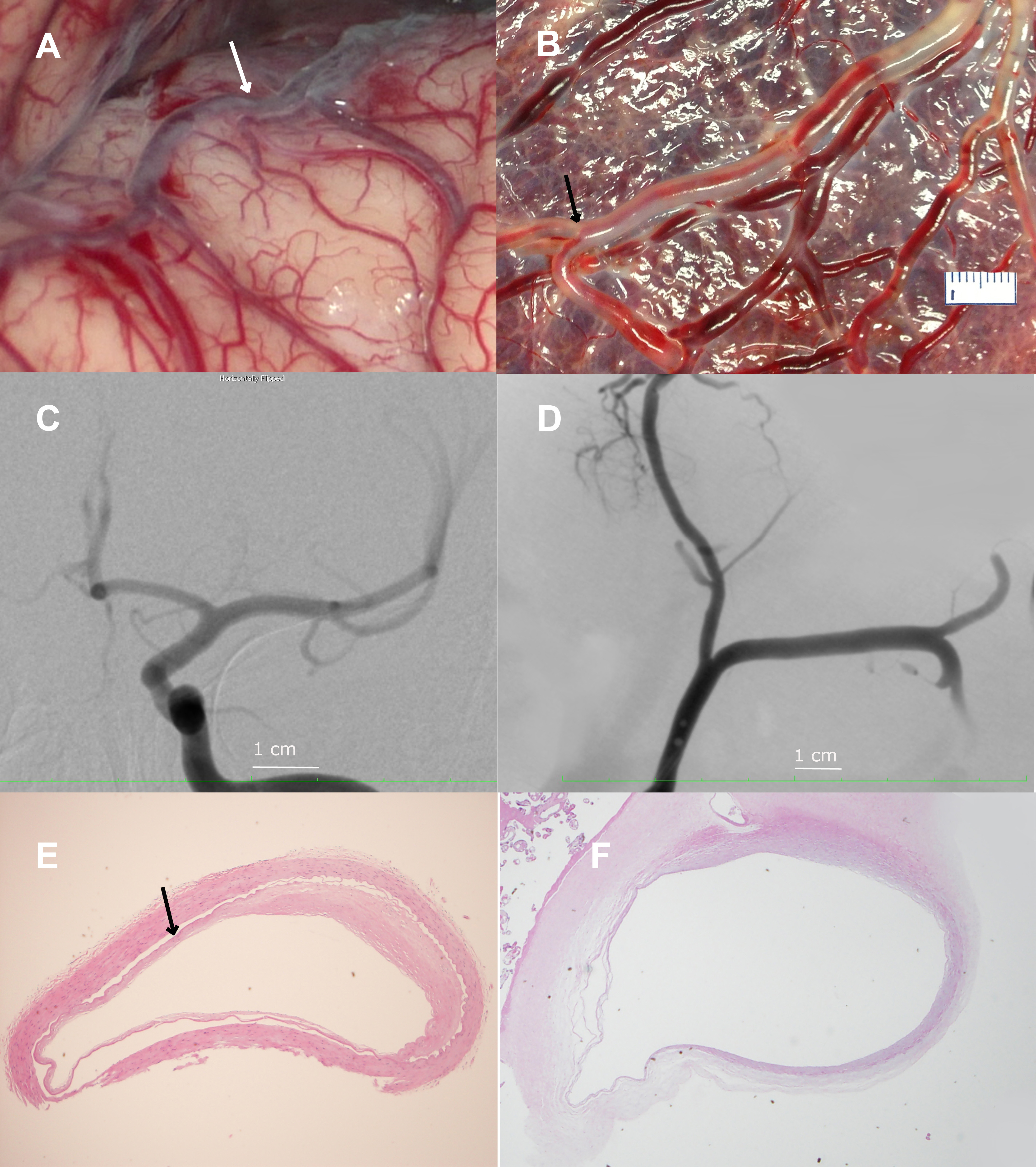
The chorional vessels are similar in size both microscopically and under floroscopic examination with contrast. Histologically, the three vascular layers are similar.
Because of the similarity of the placental vessels to the brain vesculature, our team has adopted it as a good testing model for endovascular devices and clot removal experiment. The placenta can be studied radiologically by DSA after contrast injection. Injection of flurorescent dye such as Indocargo green ICG can also be used. Under microscope mounted with 800 nm wave length flilter, the vessel can be visualized and recorded.
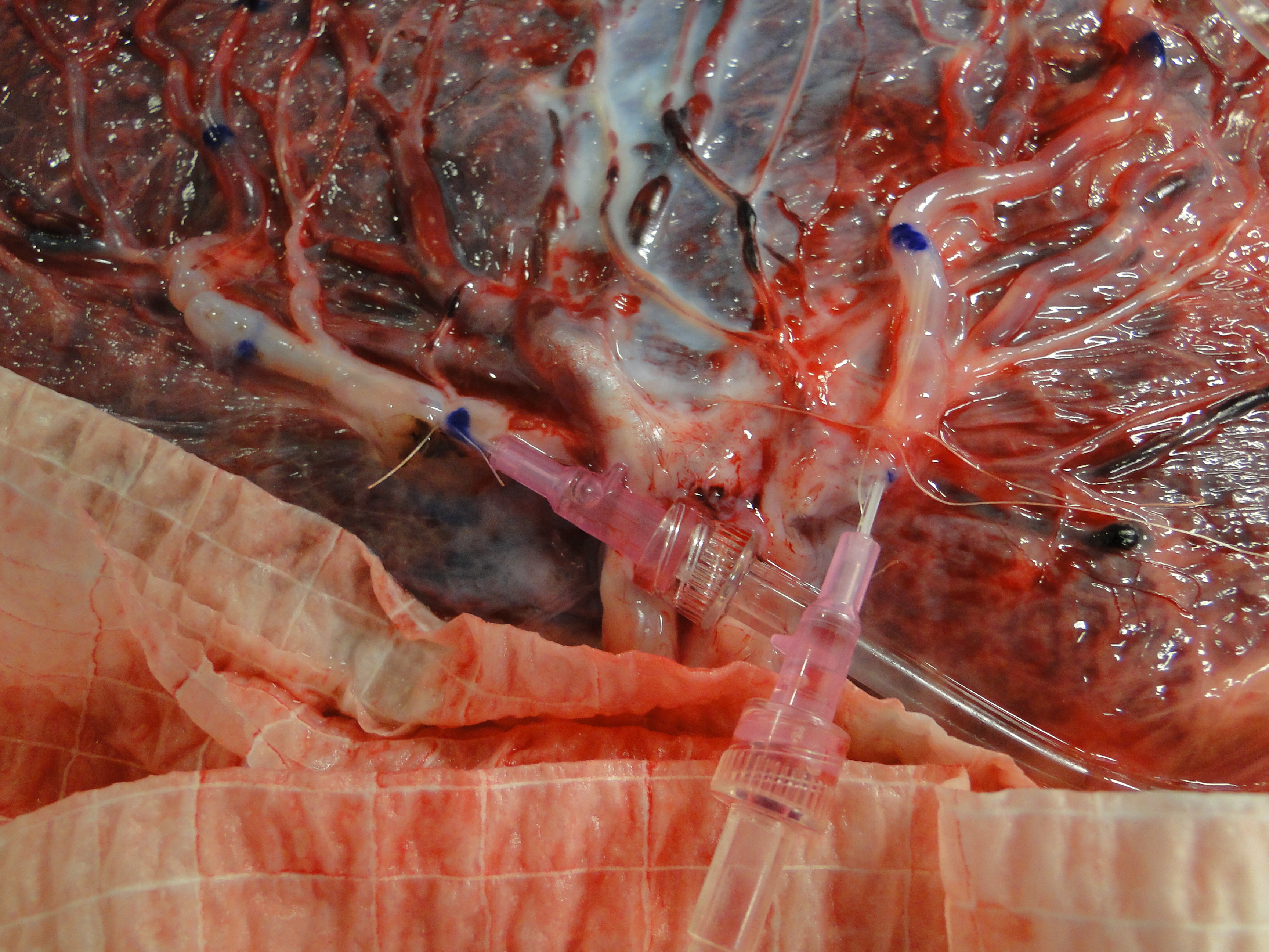
The branches of the placental arteries are catheterized and the vessels are labeled for histology.
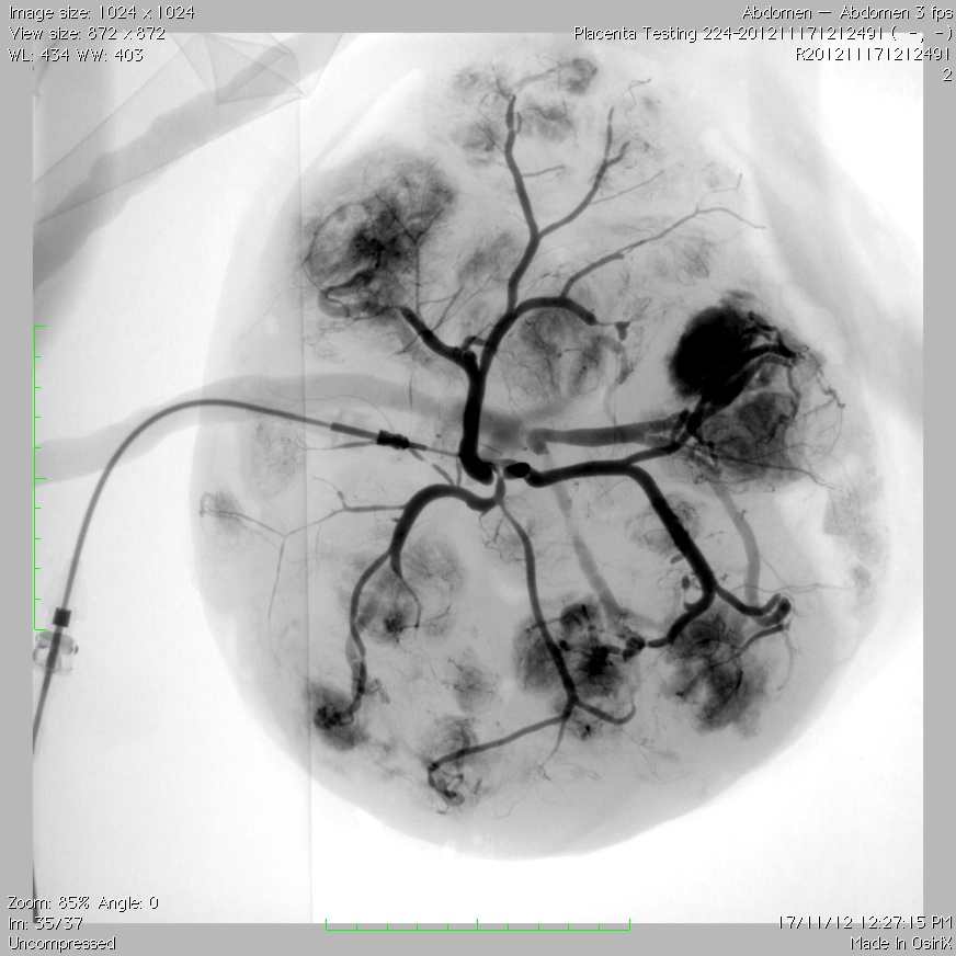
After catheterization, the vessel distribution for the placenta is studied by injection of contrast medium. DSA is taken.
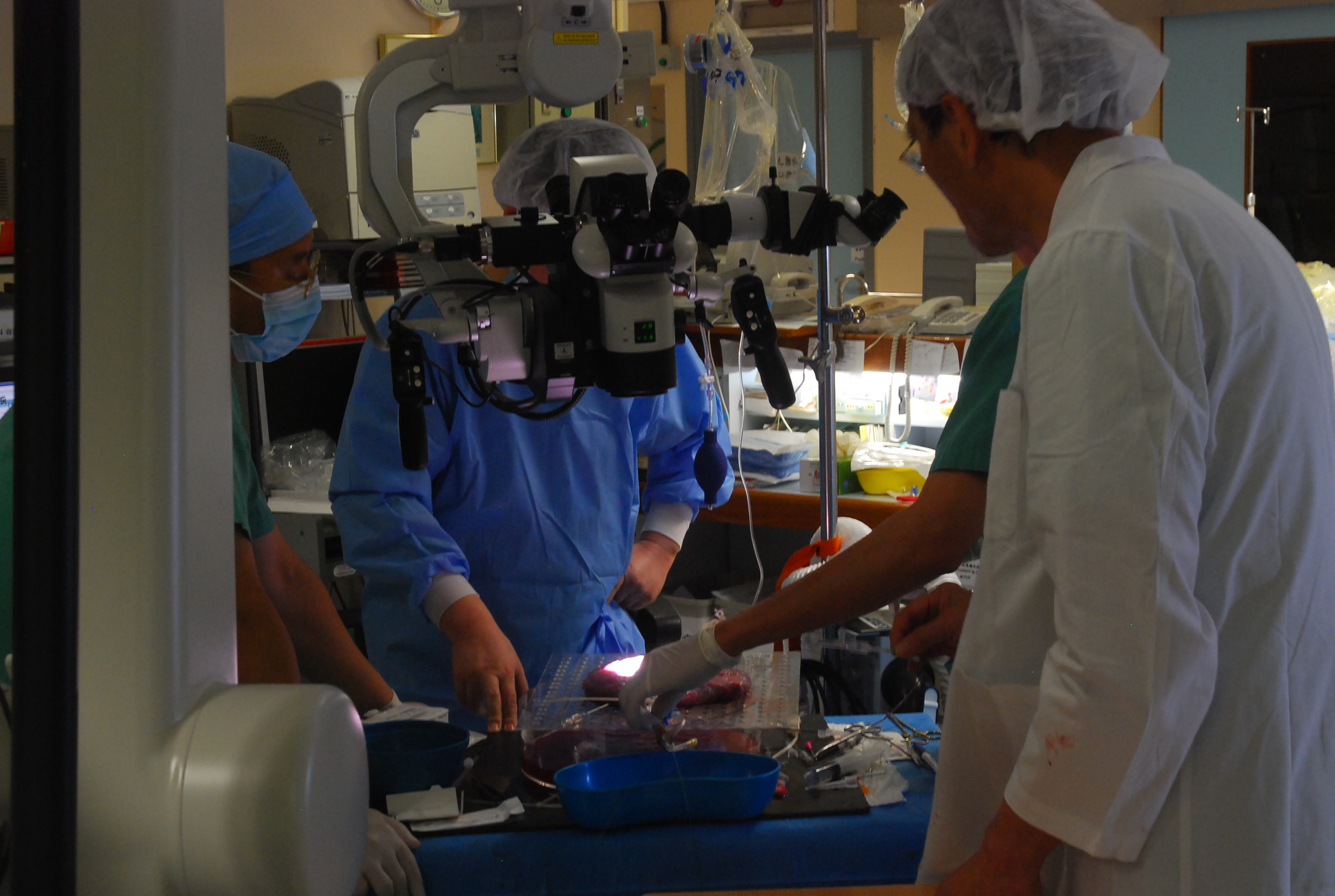
Placenta studied under surgical microscope.
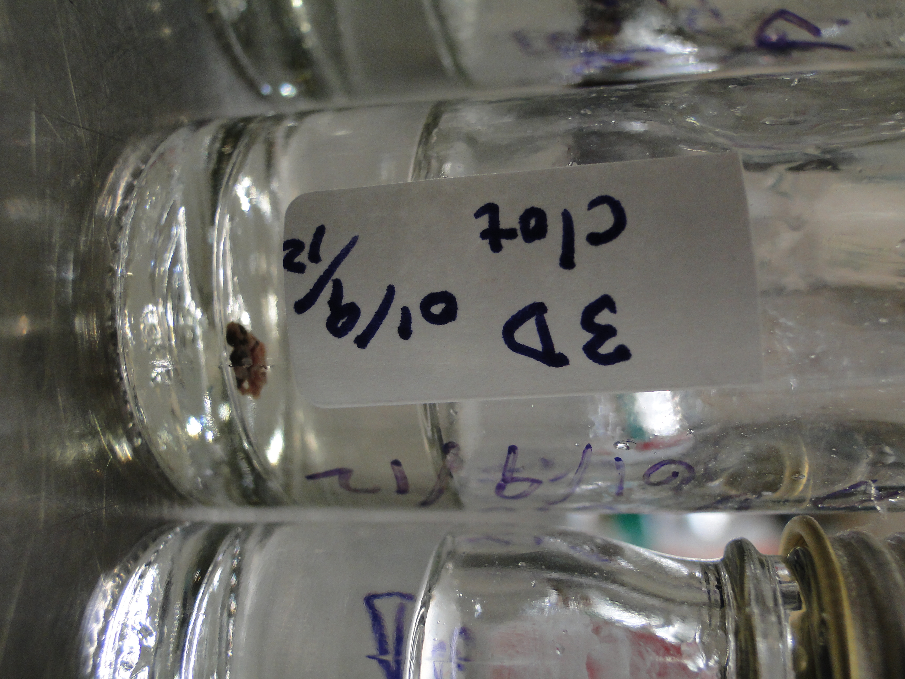
Specimen of the vessel is studied with various staining technique.